
Amygdala is the key to the expression of fear
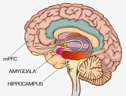
Parts of the brain involved in post-traumatic stress disorder is the amygdala, an almond-shaped region (amygdala is Greek for almond) that is key to normal expression of emotions especially fear.
Brain imaging studies revealed high activity in the amygdala region deep in the brain when subjects experience anxiety, stress or phobias.
The ventromedial prefrontal cortex (vmPFC), the hippocampus and the amygdala appeared to be altered. The vmPFC is more sophisticated part of the brain involved in less well-defined activities such as “emotional processing” and “decision making.”
However, the hippocampus region involved in memory, especially spatial memory (of places) which is a large region in the brain.
When a person encounters some environmental cue that signals danger, the information is sent to the amygdala for processing and it responds by sending out “fight or flight” responses to other parts of the brain. The vmPFC with it’s higher thinking analysed the entire situation deeply and give a logical explanation for a scenario like seeing a lion, look the lion is in an enclosure in a zoo and can’t escape so calm down and this quiet word with amygdala keep fearful responses in control.
Brain imaging studies of PTSD sufferers generally show reduced vmPFC activity and increased activity in the amygdala and produce many of the intense panic attack and anxiety symptoms that are a key feature of Post-traumatic stress disorder.
All these studies show the vital importance of addressing human psychiatric disorders with a basic scientific approach, as brain images can only show a snapshot of the current situation.
Koenig and Grafman’s clever use of the Vietnam Head injury study allowed them to ask a question that would otherwise have been difficult to answer and revealed vital new information for the development of treatments to combat PTSD.