
Heart attack risk visible by scanning blood vessels: Oxford University
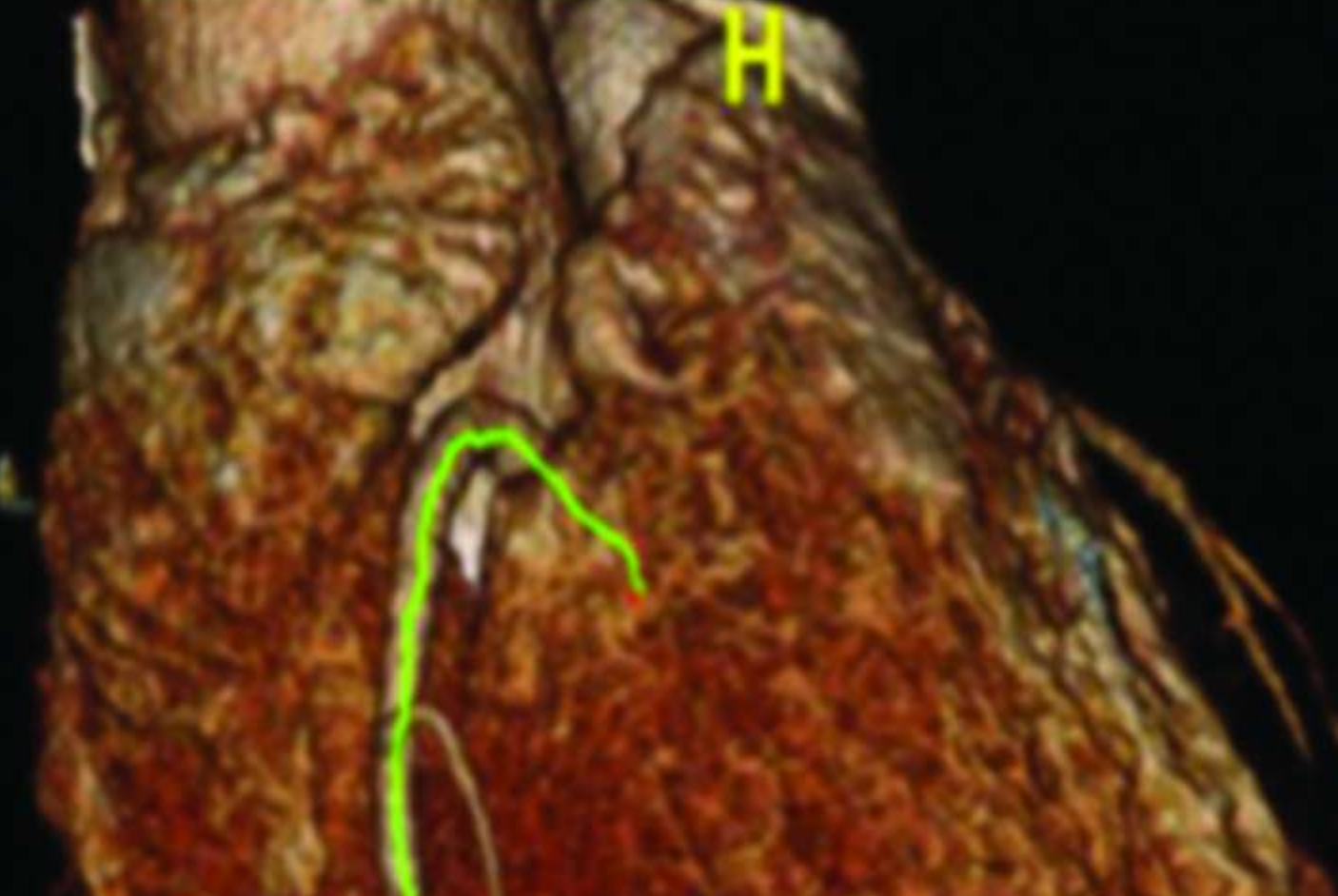
Oxford University Medical researchers have developed a sensitive test to identify people at high risk of a heart attack or stroke by scanning their blood vessels, a method known as fat attenuation index or FAI, which identifies people whose blood vessels have fatty deposits or “plaques” that are in charge of breaking off and causing a heart attack by blocking a blood vessel.
The Journal Science Translational Medicine which published their research uses new software to analyse images from computerised tomography, or CT angiograms.
Charalambos Antonlades, the Oxford project leader said ” FAI has the potential to alleviate the burden on the healthcare system by enabling resources to be concentrated on patients in greatest need of treatment”.
The Oxford team developed FAI by comparing CT images with tissue samples in 453 patients undergoing heart surgery and a further 273 patients has this technique validated. The scientist have applied for a patent and set up a spinout company, Caristo Diagnostics to commercialise the technology.